





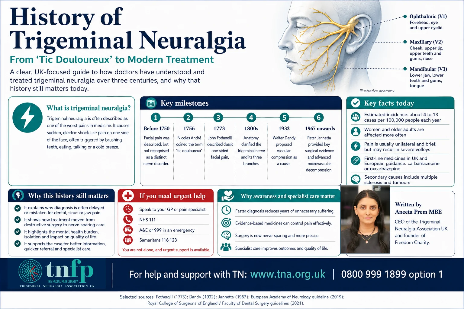
Trigeminal neuralgia has long occupied a distinctive place in the history of pain medicine. It causes sudden, severe attacks of facial pain, commonly described as sharp, shooting or electric-shock-like. The NHS describes the pain as severe and often excruciating, with attacks capable of stopping a person in their tracks.
Its reputation for exceptional severity has produced dramatic descriptions in medical history and patient advocacy. Those descriptions should not be mistaken for a scientific ranking of one painful condition against another. The clinical evidence and the effect on daily life are serious enough without sensational labels.
This history matters because the story of trigeminal neuralgia is also the story of medicine learning to distinguish nerve pain from toothache, understand the anatomy of the trigeminal nerve, recognise neurovascular compression and move from destructive operations towards treatments intended to preserve nerve function.
Before trigeminal neuralgia had a name
Descriptions resembling trigeminal neuralgia appear in medical writing stretching back centuries, although historians caution against retrospectively diagnosing every ancient account of facial or head pain. Modern historical reviews identify clearer descriptions from the seventeenth century, including accounts associated with John Locke, before the condition acquired a more recognisable medical identity in the eighteenth century.
1756: Nicolas André and “tic douloureux”
In 1756 the French surgeon Nicolas André used the term tic douloureux—literally “painful tic”—for attacks of facial pain associated with involuntary facial movement. The name survived for generations and still appears in historical medical literature.
At that time, however, physicians did not yet understand the disorder in the neurological terms used today. Treatments reflected contemporary theories of disease and could be invasive, ineffective or harmful.
1773: John Fothergill gives the classic clinical description
A decisive moment came in London in 1773, when the physician John Fothergill presented his account of what he called a painful affection of the face. His description of sudden, severe, recurrent facial pain helped establish trigeminal neuralgia as a recognisable clinical syndrome.
Fothergill’s work remains relevant because diagnosis of trigeminal neuralgia still depends heavily on the pattern of symptoms: where the pain occurs, how long attacks last, what they feel like and what triggers them. Modern imaging can help investigate causes and exclude other disorders, but the patient’s history remains central.
The trigeminal nerve comes into focus
During the nineteenth century, advances in anatomy and neurology made it possible to connect these attacks with the fifth cranial nerve—the trigeminal nerve—which carries facial sensation and has three principal divisions: ophthalmic (V1), maxillary (V2) and mandibular (V3).
This anatomical understanding helped distinguish trigeminal neuralgia from dental disease and other causes of facial pain. That distinction remains clinically important today. The NHS notes that many people with trigeminal neuralgia first seek dental care because pain is often felt in the jaw, teeth or gums.
When treatment meant damaging the nerve
Before effective medicines were available, surgeons often tried to stop pain by interrupting or destroying parts of the trigeminal nerve. Nerve section, ganglion procedures, alcohol injections and other destructive techniques developed during the nineteenth and early twentieth centuries.
These approaches could relieve pain, but at a price. Facial numbness, loss of corneal sensation and, in some cases, painful numbness known as anaesthesia dolorosa became recognised complications. The history is a reminder that pain relief and preservation of neurological function are not always the same objective.
Walter Dandy and the vascular-compression idea
A major conceptual shift came through the work of American neurosurgeon Walter Dandy. During posterior-fossa operations in the 1920s and 1930s, Dandy observed blood vessels in contact with or compressing the trigeminal nerve in patients with trigeminal neuralgia. His observations helped establish the idea that neurovascular compression could be an important mechanism in classical trigeminal neuralgia.
Dandy’s work did not immediately produce the modern operation used today, but it changed the question. Instead of asking only how to destroy pain-carrying fibres, neurosurgeons increasingly asked whether relieving pressure on the nerve might treat the disorder while preserving it.
1940s–1960s: medicines transform treatment
The arrival of anticonvulsant medicines changed the treatment landscape. Phenytoin was reported as helpful in trigeminal neuralgia in the 1940s. Carbamazepine followed and became the cornerstone of medical treatment.
Carbamazepine remains the medicine usually offered first for trigeminal neuralgia in UK guidance. Its continued importance more than half a century later is striking: despite major advances in imaging and surgery, a medicine introduced into TN treatment in the twentieth century remains central to care.
Other medicines may be considered when carbamazepine is unsuitable, ineffective or poorly tolerated, but treatment must be individualised and supervised by an appropriately qualified clinician.
Jannetta and the development of microvascular decompression
In the 1960s, Peter Jannetta used the operating microscope to develop and advance the neurovascular-compression concept. Microvascular decompression, or MVD, seeks to separate an offending blood vessel from the trigeminal nerve rather than intentionally damage the nerve.
The operation became one of the defining developments in modern trigeminal neuralgia surgery. Historical scholarship now places it within a longer line of work involving Dandy and other neurosurgeons rather than attributing the entire concept to a single moment or individual.
MVD may provide substantial and sometimes long-lasting pain relief for appropriately selected patients, but it is major intracranial surgery and carries important risks. No treatment works for everyone.
Percutaneous procedures and radiosurgery
Other procedures developed alongside MVD. Percutaneous treatments—including radiofrequency lesioning, glycerol injection and balloon compression—aim to interrupt pain transmission through controlled injury to trigeminal fibres. Stereotactic radiosurgery uses focused radiation to target the nerve without an open operation.
Each approach involves different trade-offs between invasiveness, durability, numbness and other complications. The modern principle should be informed choice: patients need clear information about benefits, uncertainties and risks rather than a single procedure being presented as universally best.
Modern understanding: classical, secondary and idiopathic TN
Today, trigeminal neuralgia is not treated as one uniform disease. Contemporary classification distinguishes classical trigeminal neuralgia, associated with neurovascular compression producing morphological change in the nerve; secondary trigeminal neuralgia, caused by another neurological disorder such as multiple sclerosis or a space-occupying lesion; and idiopathic trigeminal neuralgia, where no clear cause meeting those categories is established.
This distinction matters. It affects investigation, discussion of treatment and the interpretation of imaging. A blood vessel touching the trigeminal nerve on a scan is not, by itself, enough to explain every case of facial pain. Clinical assessment remains essential.
The part of the history medicine neglected
For much of the recorded history of trigeminal neuralgia, medical writing concentrated on anatomy, medicines and operations. Patients were often described through procedures rather than through the effect of pain on their lives.
That is changing. Severe and unpredictable facial pain can affect eating, speaking, brushing teeth, washing, work, sleep, relationships, confidence and mental health. The NHS recognises that trigeminal neuralgia can significantly affect quality of life and may contribute to isolation and depression.
Trigeminal neuralgia does not need frightening nicknames to be taken seriously. Its documented clinical severity and human impact are powerful enough.
Why the history still matters
The history of trigeminal neuralgia reveals several recurring failures. Patients can still be misdirected towards dental treatment before the neurological pattern is recognised. Diagnosis can still take too long. Treatment can still involve difficult compromises between pain relief, side effects and neurological function.
But the history also shows progress. Medicine moved from vague descriptions of facial agony to a recognisable syndrome; from destructive nerve surgery to nerve-sparing decompression for selected patients; from almost no effective medicines to established pharmacological treatment; and from viewing pain only as an anatomical event to recognising its effect on the whole person.
For me, that history has another lesson. Patient testimony is not the opposite of medical evidence. It is often where the unanswered question first becomes visible.
The responsibility of modern medicine is not to rank suffering. It is to recognise the pattern, diagnose accurately, explain uncertainty, treat intelligently and make sure nobody living with severe facial pain becomes invisible.
About Aneeta Prem MBE
Aneeta Prem MBE is Chief Executive of Trigeminal Neuralgia Association UK and lives with bilateral trigeminal neuralgia. Her first severe facial-pain attack was in September 2009. She writes about patient experience, diagnosis, facial pain, safeguarding and access to care.
Sources and further reading
- NHS: Trigeminal neuralgia
- NHS: Trigeminal neuralgia symptoms
- NHS: Diagnosis
- NHS: Treatment
- Historical characterisation of trigeminal neuralgia
- Historical evolution of microvascular decompression
- Historical perspectives on diagnosis and treatment
Related on Aneeta.com
Substantively reviewed and updated: 21 August 2026.